
INTRA-ORAL BASAL CELL CARCINOMA- AN IMMUNOHISTOCHEMICAL INTERPRETATION USING BER–EP4
Abstract
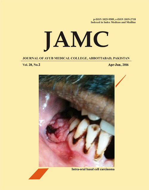
A 33 year old male presented with a painful non–healing growth of gingiva from eight months. Intra–oral examination revealed an indurated and erythematous lesion on gingiva with relation to tooth #29 to missing #30 measuring 2×1 cm. (Figure-1) Radiographic examination revealed a bone loss with relation to tooth #29 and #30. An incisional biopsy was performed and final diagnosis of peripheral ameloblastoma was given. At the time of excisional biopsy patient came up with increased swelling which remained ulcerated and the site of incisional biopsy was not healed. Histopathological examination of excisional biopsy showed numerous islands of basaloid cells budding off from the basal cell layer of epithelium. (Figure-2) These islands comprised of numerous apoptotic cells and mitotic figures. The peripheral cells of the islands demonstrated a palisaded arrangement. These features were similar to peripheral ameloblastoma, (PA)1 we decided to do immuno-histochemical (IHC) staining for Ber-EP4 (to rule out PA). IHC stain for Ber-EP4 showed positive staining for the lesional cells of invading islands. (Figure-3). Based on IHC profile, a final diagnosis of mucosal basal cell carcinoma was rendered. One year follow up of the patient is uneventful.Intra oral Basal cell carcinoma (IOBCC) is a rare and controversial entity.2 Because of very close resemblance with PA it is often misdiagnosed on histopathological grounds, as in the current case. Ber-EP4 is a monoclonal antibody against cell surface glycoproteins (34 and 39 kDa) of most epithelial cells that reacts with epithelial tumours, with the exception of superficial layers of squamous epithelia.3 The differentiation of IOBCC and PA is very important as IOBCC is a malignant and locally aggressive tumour. Ber EP4 is a reliable marker to differentiate IOBCC from PA. This case highlights the importance of immunohistochemistry in diagnosis.References
Bajpai M, Pardhe N. Peripheral Ameloblastoma with Mixed Histological Patterns. Cukurova Med J 2015;40:151–5.
Tellechea O, Reis JP, Domingues JC, Baptista AP. Monoclonal antibody Ber EP4 distinguishes basal-cell carcinoma from squamous- cell carcinoma of the skin. Am J Dermatopathol 1993;15(5):452–5.
Latza U, Niedobitek G, Schwarting R, Nekarda H, Stein H. Ber- EP4: new monoclonal antibody which distinguishes epithelia from mesothelial. J Clin Pathol 1990; 43(3):213–9.
Published
Issue
Section
License
Journal of Ayub Medical College, Abbottabad is an OPEN ACCESS JOURNAL which means that all content is FREELY available without charge to all users whether registered with the journal or not. The work published by J Ayub Med Coll Abbottabad is licensed and distributed under the creative commons License CC BY ND Attribution-NoDerivs. Material printed in this journal is OPEN to access, and are FREE for use in academic and research work with proper citation. J Ayub Med Coll Abbottabad accepts only original material for publication with the understanding that except for abstracts, no part of the data has been published or will be submitted for publication elsewhere before appearing in J Ayub Med Coll Abbottabad. The Editorial Board of J Ayub Med Coll Abbottabad makes every effort to ensure the accuracy and authenticity of material printed in J Ayub Med Coll Abbottabad. However, conclusions and statements expressed are views of the authors and do not reflect the opinion/policy of J Ayub Med Coll Abbottabad or the Editorial Board.
USERS are allowed to read, download, copy, distribute, print, search, or link to the full texts of the articles, or use them for any other lawful purpose, without asking prior permission from the publisher or the author. This is in accordance with the BOAI definition of open access.
AUTHORS retain the rights of free downloading/unlimited e-print of full text and sharing/disseminating the article without any restriction, by any means including twitter, scholarly collaboration networks such as ResearchGate, Academia.eu, and social media sites such as Twitter, LinkedIn, Google Scholar and any other professional or academic networking site.


