
NEURILEMMOMA OF EXTERNAL EAR, CONFIRMED BY IMMUNOHISTOCHEMISTRY–A TELEPATHOLOGICAL COMMUNICATION BETWEEN CYPRUS AND INDIA
Abstract
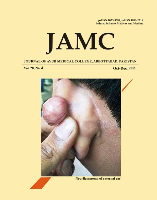
A 27 year old Cypriot male presented to the Hippocrateon hospital of dermatology for the evaluation of a painless swelling of external ear persisted from 1 year duration. Initially it was small but gradually reached to its current size.Clinical examination revealed a round swelling on pinna measuring about 3×3 cm in dimension. The overlying skin was slightly reddish in colour without any ulceration. On palpation the swelling was found to be movable. (Figure-1). The cervical lymph nodes were non–palpable. Fine needle Aspiration cytology was done but it was not conclusive. Based on the site and clinical feature, provisional diagnosis of benign adnexal tumour was made. Finally an operation was planned and the lesion was excised conservatively under anaesthesia.The Microscopic examination using Hematoxylin and Eosin stains showed numerous cords of cells that had either short, fusiform or rounded shapes, histologically identical to shwann cells separated by fibrous septa. Intra – nodular cells, at few areas exhibited a typical Antoni A appearance with a palisaded arrangement; however few areas showed a loss of palisading signified Antoni B type of arrangement. (Figure-2) An area of haemorrhage showed a collection of numerous extra - vasated RBCs. Few areas showed high vascularity with multiple dilated blood vessels surrounded by endothelial cells.The diagnosis of Neurilemmoma was made on the basis of Histological pictures. We approached Dr Manas Bajpai (Oral and Maxillofacial Pathologist, India) for expert comments and his opinion about the diagnosis and follow up of the patient through email and telephonic conversation. Dr Bajpai confirmed our diagnosis, he further suggested an Immuno-histochemical staining using antibodies to S-100 proteins. Immuno-histochemical staining of the specimen for S -100 marker was carried out using immuno simple stain kit (Athens, Greece)The immune-histochemical staining revealed a strong expression of S – 100 markers for the cell. (Figure -3) Hence it confirmed the diagnosis of Neurilemmoma.Neurilemmoma/Scwannoma is a benign peripheral nerve sheath tumour.1 clinically presents as a slow growing mass. Definite diagnosis should be based on the histological and immune-histochemical findings. Typically, histological analysis demonstrates that a schwannoma is composed of S-100 protein positive Schwann cells arranged in 2 growth patterns, namely Antoni A and B.2 Very few cases of neurilemmoma of external ear has been reported in the literature.The present case describes a rare lesion of external ear; also it highlights the importance of tele-pathology in routine diagnosis and the utility of immunohistochemistry in final diagnosis.Tele-pathology is the practice of pathology at a distance. It uses telecommunications technology to facilitate the transfer of image-rich pathology data between distant locations for the purposes of diagnosis, education, and research. 3 The present case was diagnosed by the communication between a dermatologist of Nicosia (Cyprus) and Oral and Maxillofacial Pathologist of Jaipur (India)..References
Kaiserling E, Geerts ML. Tumour of Wagner-Meissner touch corpuscles. Wagner-Meissner neurilemmoma. Virchows Arch A Pathol Anat Histopathol 1986;409(2):241–50.
van Zuuren EJ, Posma AN. Diffuse neurofibroma on the lower back. J Am Acad Dermatol 2003;48(6):938–40.
Kumar S. Telepathology: An Audit. In: Kumar S, Dunn BE, editors. Telepathology. Berlin, Heidelberg: Springer Berlin Heidelberg; 2009. p. 225–8.
Published
Issue
Section
License
Journal of Ayub Medical College, Abbottabad is an OPEN ACCESS JOURNAL which means that all content is FREELY available without charge to all users whether registered with the journal or not. The work published by J Ayub Med Coll Abbottabad is licensed and distributed under the creative commons License CC BY ND Attribution-NoDerivs. Material printed in this journal is OPEN to access, and are FREE for use in academic and research work with proper citation. J Ayub Med Coll Abbottabad accepts only original material for publication with the understanding that except for abstracts, no part of the data has been published or will be submitted for publication elsewhere before appearing in J Ayub Med Coll Abbottabad. The Editorial Board of J Ayub Med Coll Abbottabad makes every effort to ensure the accuracy and authenticity of material printed in J Ayub Med Coll Abbottabad. However, conclusions and statements expressed are views of the authors and do not reflect the opinion/policy of J Ayub Med Coll Abbottabad or the Editorial Board.
USERS are allowed to read, download, copy, distribute, print, search, or link to the full texts of the articles, or use them for any other lawful purpose, without asking prior permission from the publisher or the author. This is in accordance with the BOAI definition of open access.
AUTHORS retain the rights of free downloading/unlimited e-print of full text and sharing/disseminating the article without any restriction, by any means including twitter, scholarly collaboration networks such as ResearchGate, Academia.eu, and social media sites such as Twitter, LinkedIn, Google Scholar and any other professional or academic networking site.


