
WARTHIN'S TUMOUR OF FLOOR OF THE MOUTH - CORRELATION OF FNAC AND HISTOPATHOOGICAL FINDINGS
Abstract
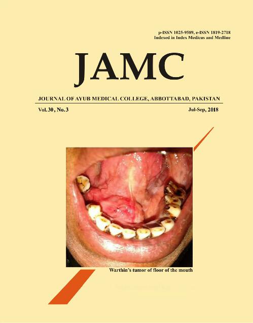
A 43-year-old male presented to our institute with the chief complain of a painless swelling of floor of the mouth from 8 months. The swelling was initially small and gradually reached its current size. Personal history of the patient revealed the he had been consuming 8-10 cigarettes daily from 15 years. The family history and past medical history of the patient was non - contributory to the presenting symptom. Intra-oral examination revealed a solitary dome shaped soft and fluctuant tissue growth of the right side of the floor of the mouth extending from lingual mucosa with relation to the tooth #41 to tooth #44 measuring about 3×2 cm. The colour of the overlying mucosa was similar to the adjacent mucosa without any sign of ulceration and discharge. [Figure-1a] FNAC was performed under aseptic condition. Smears were wet fixed immediately in ether-alcohol fixative and stained by Papanicolaou stain. The microscopic examination revealed a moderately cellular field comprised of single layered cohesive sheets of epithelial cells with focal collection of lymphocytes in a mucoid background. [Figure-1b] The epithelial cells were showing oncocytic pattern with densely eosinophilic granular cytoplasm with round nuclei. [Figure-1c] Based on cytopathological findings an initial impression of WT was made. The surgical excision of the lesion was done under general anaesthesia and the excised tissue was sent for histopathological evaluation.
The histopathological examination of haematoxylin and eosin stained soft tissue section revealed cystic spaces densely filled with eosinophilic coagulum and surrounded by bilayered oncocytic epithelium arranged in papillary fronds. The inner cell layer was tall columnar and outer layer was oncocytic. These papillary fronds were surrounded by dense lymphocytic infiltrates. [Figure 1d] Based on histopathology a final diagnosis of Warthin's tumour was given.
Warthin's tumours (WT) are most commonly found in parotid gland.1-3 They are the second most common tumours of parotid after pleomorphic adenoma.2 WT occurring in minor salivary gland of floor of the mouth is an exceedingly rare phenomenon. An exhaustive literature review has revealed a single published case of WT of floor of the mouth; hence the present case is being only the second case.3
The occurrence of Warthin's tumour in minor salivary glands is extremely rare and the incidence was reported between 0.1-1.2%.1,2 The Incidence of WT commonly appears between ages of 58 and 70 years and can arise in adults; WT is more common in men than in women. Previous studies have shown a close association of WT with smoking. In present case also the patient was a chronic smoker.1-3 The accuracy of FNAC in diagnosis of salivary gland tumours is quite debatable among clinicians and cytopathologist. FNAC in WT may simulate many other conditions including oncocytoma, oncocytic carcinoma and lymphoepithellial cyst.1A proper care and knowledge pertaining to the limitations of cytology in diagnosing WT is needed.
References
Warthin AS. Papillary cystadenoma lymphomatosum. A rare teratoid of the parotid region. J Cancer Res 1929;13(2):116-25.
Faur A, Lazar E, Cornianu M, Dema A, Vidita CG, Galuscan A. Warthin tumor: a curious entity-- case reports and review of literature. Rom J Morphol Embryol 2009;50(2):269-73.
Hatch RL, Shah S. Warthin tumour: a common, benign tumour presenting as a highly suspicious mass. J Am Board Fam Pract 2005;18(4):320-2.
Published
How to Cite
Issue
Section
License
Journal of Ayub Medical College, Abbottabad is an OPEN ACCESS JOURNAL which means that all content is FREELY available without charge to all users whether registered with the journal or not. The work published by J Ayub Med Coll Abbottabad is licensed and distributed under the creative commons License CC BY ND Attribution-NoDerivs. Material printed in this journal is OPEN to access, and are FREE for use in academic and research work with proper citation. J Ayub Med Coll Abbottabad accepts only original material for publication with the understanding that except for abstracts, no part of the data has been published or will be submitted for publication elsewhere before appearing in J Ayub Med Coll Abbottabad. The Editorial Board of J Ayub Med Coll Abbottabad makes every effort to ensure the accuracy and authenticity of material printed in J Ayub Med Coll Abbottabad. However, conclusions and statements expressed are views of the authors and do not reflect the opinion/policy of J Ayub Med Coll Abbottabad or the Editorial Board.
USERS are allowed to read, download, copy, distribute, print, search, or link to the full texts of the articles, or use them for any other lawful purpose, without asking prior permission from the publisher or the author. This is in accordance with the BOAI definition of open access.
AUTHORS retain the rights of free downloading/unlimited e-print of full text and sharing/disseminating the article without any restriction, by any means including twitter, scholarly collaboration networks such as ResearchGate, Academia.eu, and social media sites such as Twitter, LinkedIn, Google Scholar and any other professional or academic networking site.


