
OPTIC DISC MELANOCYTOMA; A RARE ENTITY
Abstract
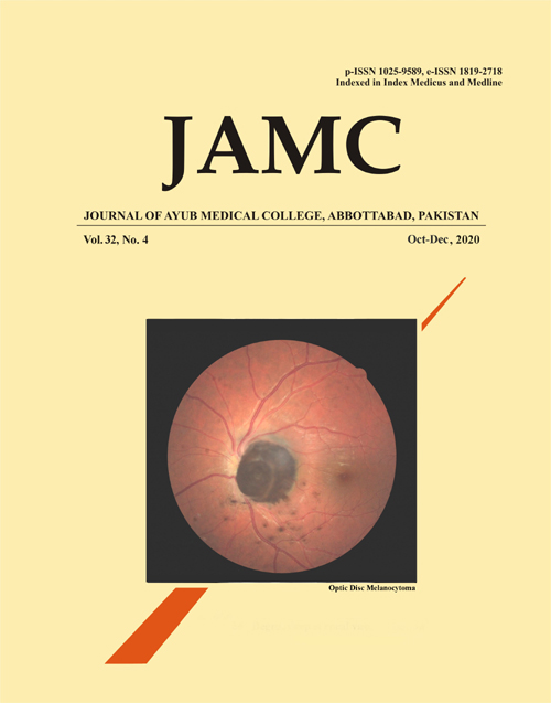
A young male presented with complaints of disturbance of vision and floaters for the past five years with best corrected visual acuity of 6/6 in both eyes. Ophthalmic examination of the anterior segment was normal in both eyes. There was a relative afferent papillary defect in the right eye. Fundoscopic examination of the left eye showed a raised, 3-disc diameter (DD) dark brown lesion arising from the optic disc. Ophthalmic investigations were performed including fundus photos, A-scan, B-scan, Auto-fluorescence, OCT, and FFA. Findings were consistent with those of optic disc melanocytoma. The patient is now kept on close three-monthly follow-up. To the best of our knowledge, there are no published reports from Pakistan so far.
Keywords: Optic Disc Melanocytoma; Melanocytic Lesion; Choroidal MelanomaReferences
Harbour JW, Paez-Escamilla M, Cai L, Walter SD, Augsburger JJ, Correa ZM. Are risk factors for growth of choroidal nevi associated with malignant transformation? Assessment with a validated genomic biomarker. Am J Ophthalmol 2019;197:168-79.
Pereira A, Thomas S, Yadav NK, Venkatesh R. Multicolor imaging of optic disc melanocytoma. Indian journal of ophthalmology. 2019 Dec;67(12):2056.
Mansour AM, Zimmerman LO, La Piana FG, Beauchamp GR. Clinicopathological findings in a growing optic nerve melanocytoma. Br J Ophthalmol 1989;73(6):410-5.
Baartman BJ, Ahmad B, Srivastava S, Jones S, Singh AD. Melanocytoma or juxtapapillary melanoma?. Retinal Cases and Brief Reports. 2019 Jan 1;13(1):15-7.
Attiku Y, Rishi P, Bassi S. Coexisting Optic Disc Melanocytoma and Pituitary Adenoma. Ocul Oncol Pathol 2019;5(5):319-22.
Herwig-Carl MC, Loeffler KU, Grossniklaus HE. Melanocytoma of the conjunctiva: clinicopathologic features of three cases. Ocular oncology and pathology. 2019;5(4):290-7.
Al-Rashaed S, Abboud EB, Nowilaty SR. Characteristics of optic disc melanocytomas presenting with visual dysfunction. Middle East Afr J Ophthalmol 2010;17(3):242-5.
Kita Y, Hollό G, Murai A, Kita R, Hirakata A. Optical coherence tomography angiography findings of an optic disc melanocytoma in a glaucoma eye. International Ophthalmology. 2019 Mar 15;39(3):677-82.
Carnevali A, Querques L, Zucchiatti I, Scorcia V, Bandello F, Querques G. Optical coherence tomography angiography features in melanocytoma of the optic nerve. Ophthalmic Surg Lasers Imaging Retina 2017;48(4):364-6.
Guerra RLL, Marback EF, Silva IS, Maia Jr OD, Marback RL. Autofluorescência e tomografia de coerência óptica de domÃnio espectral do melanocitoma do disco óptico. Arq Bras Oftalmol 2014;77(6):400-2.
Zhang P, Hui YN, Xu WQ, Zhang ZF, Wang HY, Sun DJ, et al. Infrared autofluorescence, short-wave autofluorescence and spectral-domain optical coherence tomography of optic disk melanocytomas. Int J Ophthalmol 2016;9(5):713-6.
Shields JA, Demirci H, Mashayekhi A, Eagle Jr RC, Shields CL. Melanocytoma of the optic disk: A review. Indian J Ophthalmol 2019;67(12):1949-58.
Downloads
Published
How to Cite
Issue
Section
License
Journal of Ayub Medical College, Abbottabad is an OPEN ACCESS JOURNAL which means that all content is FREELY available without charge to all users whether registered with the journal or not. The work published by J Ayub Med Coll Abbottabad is licensed and distributed under the creative commons License CC BY ND Attribution-NoDerivs. Material printed in this journal is OPEN to access, and are FREE for use in academic and research work with proper citation. J Ayub Med Coll Abbottabad accepts only original material for publication with the understanding that except for abstracts, no part of the data has been published or will be submitted for publication elsewhere before appearing in J Ayub Med Coll Abbottabad. The Editorial Board of J Ayub Med Coll Abbottabad makes every effort to ensure the accuracy and authenticity of material printed in J Ayub Med Coll Abbottabad. However, conclusions and statements expressed are views of the authors and do not reflect the opinion/policy of J Ayub Med Coll Abbottabad or the Editorial Board.
USERS are allowed to read, download, copy, distribute, print, search, or link to the full texts of the articles, or use them for any other lawful purpose, without asking prior permission from the publisher or the author. This is in accordance with the BOAI definition of open access.
AUTHORS retain the rights of free downloading/unlimited e-print of full text and sharing/disseminating the article without any restriction, by any means including twitter, scholarly collaboration networks such as ResearchGate, Academia.eu, and social media sites such as Twitter, LinkedIn, Google Scholar and any other professional or academic networking site.


