
INTRAORAL VERRUCA VULGARIS
Abstract
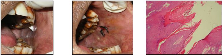
A 58-year-old male visited to the Department of Oral medicine and radiology in Indraprastha Dental Hospital (India) with chief complaints of a white lesion in the left buccal mucosa since last 4 month which was gradually increasing in size. His past medical history was non-contributory and similar lesions are not present among his other family members. On intraoral examination, a solitary proliferative verrucous growth over the left buccal mucosa was noted. The lesion was exophytic and sessile in nature, approximately 1×1 cm in size, with well-defined margins. Surface of the lesion was irregular with finger-like projections. The colour was white, non-tender and soft in consistency [Figure-1] Multiple differential diagnosis was given as squamous papilloma, condylomata accuminatum, keratoacanthoma, exophytic verrucous carcinoma, focal epithelial hyperplasia, verruciform xanthoma. So, a biopsy was decided. The lesion was excised completely under local anaesthesia and sutured. [Figure-2] The excised biopsy sample was sent for histopathological examination. [Figure3] Under hemotoxin and eosin staining showed superficial layers of the epithelium demonstrate koilocytotic changes. There is polypoid mass with an epithelium displaying acanthosis, and papillomatosis. There was proliferation of hyperkeratotic stratified squamous epithelium arranged into pointed projections with connective tissue cores. Elongated rete ridges tend to converge toward the center of the lesion, producing a ‘cupping’ effect. This leads to final diagnosis of Verruca Vulgaris of buccal mucosa. Oral verruca vulgaris is caused by human papillomavirus (HPV) mostly by type 6 and 11 infection.1 Verruca vulgaris most frequently occurs on the fingers, toes, soles, and dorsal surfaces of hands and is mostly asymptomatic. It’s rarely seen in oral cavity. Viral products stimulate cell growth in the basal layer that leads to formation of a wart which has malignant potential due HPV infection.2 Varieties of verrucous lesions affect oral mucosa which can be reactive. It commonly observed skin growths in childhood with equal gender predilection. Intraoral warts can occur at any age with equal incidence in both genders but are most commonly seen in the third to fifth decade. Medication that can be advised are cimetidine, levamisole, retinoids, immunomodulator. It can also be removed by surgical excision or laser.3References
Major T, Szarka K, Sziklai I, Gergely L, Czeglédy J. The characteristics of human papillomavirus DNA in head and neck cancers and papillomas. J Clin Pathol 2005;58(1):51–5.
Praetorius F. HPV-associated diseases of oral mucosa. Clin Dermatol 1997;15(3):399–413.
Parsad D, Pandhi R, Juneja A, Negi KS. Cimetidine and levamisole versus cimetidine alone for recalcitrant warts in children. Pediatr Dermatol 2001;18(4):349–52.
Published
Issue
Section
License
Journal of Ayub Medical College, Abbottabad is an OPEN ACCESS JOURNAL which means that all content is FREELY available without charge to all users whether registered with the journal or not. The work published by J Ayub Med Coll Abbottabad is licensed and distributed under the creative commons License CC BY ND Attribution-NoDerivs. Material printed in this journal is OPEN to access, and are FREE for use in academic and research work with proper citation. J Ayub Med Coll Abbottabad accepts only original material for publication with the understanding that except for abstracts, no part of the data has been published or will be submitted for publication elsewhere before appearing in J Ayub Med Coll Abbottabad. The Editorial Board of J Ayub Med Coll Abbottabad makes every effort to ensure the accuracy and authenticity of material printed in J Ayub Med Coll Abbottabad. However, conclusions and statements expressed are views of the authors and do not reflect the opinion/policy of J Ayub Med Coll Abbottabad or the Editorial Board.
USERS are allowed to read, download, copy, distribute, print, search, or link to the full texts of the articles, or use them for any other lawful purpose, without asking prior permission from the publisher or the author. This is in accordance with the BOAI definition of open access.
AUTHORS retain the rights of free downloading/unlimited e-print of full text and sharing/disseminating the article without any restriction, by any means including twitter, scholarly collaboration networks such as ResearchGate, Academia.eu, and social media sites such as Twitter, LinkedIn, Google Scholar and any other professional or academic networking site.


