
CROSSED CEREBELLAR DIASCHISIS IN A CHILD WITH STATUS EPILEPTICUS: AN UNUSUAL PRESENTATION
Abstract
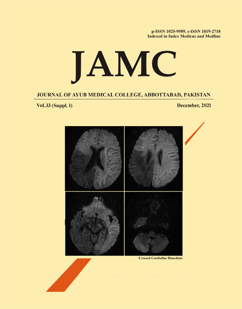
Crossed Cerebellar Diaschisis (CCD) describes a depression of oxidative metabolism and blood flow in the cerebellum secondary to a supratentorial lesion in the contralateral cerebral hemisphere. The pathophysiology is not clear but appears to be caused by abnormal neuronal connection of the primary to the remote site. The diagnosis is usually done using positron emission tomography (PET) and single-photon emission CT (SPECT) scans. Almost all the reported cases of CCD are caused by acute ischemic stroke in adults. Hence, CCD secondary to status epilepticus, extremely rare and there is limited literature available on it. This is important because it's findings can easily be confused with acute ischemic stroke and similar concurrent diseases. Correct diagnosis can also help localize the cause of the seizures and significantly influence surgical decisions. We present a case of CCD in a child with status epilepticus using MRI of the brain with DWI.
References
Han S, Wang X, Xu K, Hu C. Crossed Cerebellar Diaschisis: Three Case Reports Imaging Using a Tri-Modality PET/CT-MR System. Medicine (Baltimore) 2016;95(2):e2526.
Zaidi SA, Haq MAu, Bindman D, Mathur S. Crossed cerebellar diaschisis: a radiological finding in status epilepticus not to miss. BMJ Case Rep 2013;2013:bcr2013200478.
Katramados AM, Burdette D, Patel SC, Schultz LR, Gaddam S, Mitsias PD. Periictal diffusion abnormalities of the thalamus in partial status epilepticus. Epilepsia 2009;50(2):265-75.
Sin DS, Kim MH, Park SA, Joo MC, Kim MS. Crossed Cerebellar Diaschisis: Risk Factors and Correlation to Functional Recovery in Intracerebral Hemorrhage. Ann Rehabil Med 2018;42(1):8-17.
Oster J, Doherty C, Grant PE, Simon M, Cole AJ. Diffusion-weighted imaging abnormalities in the splenium after seizures. Epilepsia 2003;44(6):852-4.
Chakravarty A. MR evaluation of crossed and uncrossed cerebral-cerebellar diaschisis. Acta Neurol Scand 2003;108(1):60-5.
Lansberg MG, O'Brien MW, Norbash AM, Moseley ME, Morrell M, Albers GW. MRI abnormalities associated with partial status epilepticus. Neurology 1999;52(5):1021-7.
Doherty CP, Cole AJ, Grant PE, Fischman A, Dooling E, Hoch DB, et al. Multimodal longitudinal imaging of focal status epilepticus. Can J Neurol Sci 2004;31(2):276-81.
Grant PE, He J, Halpern EF, Wu O, Schaefer PW, Schwamm LH, et al. Frequency and clinical context of decreased apparent diffusion coefficient reversal in the human brain. Radiology 2001;221(1):43-50.
Kamalian S, Lev MH. Stroke Imaging. Radiol Clin N Am 2019;57(4):717-32.
Cole AJ. Status epilepticus and periictal imaging. Epilepsia. 2004;45(Suppl 4):72-7.
Downloads
Published
How to Cite
Issue
Section
License
Journal of Ayub Medical College, Abbottabad is an OPEN ACCESS JOURNAL which means that all content is FREELY available without charge to all users whether registered with the journal or not. The work published by J Ayub Med Coll Abbottabad is licensed and distributed under the creative commons License CC BY ND Attribution-NoDerivs. Material printed in this journal is OPEN to access, and are FREE for use in academic and research work with proper citation. J Ayub Med Coll Abbottabad accepts only original material for publication with the understanding that except for abstracts, no part of the data has been published or will be submitted for publication elsewhere before appearing in J Ayub Med Coll Abbottabad. The Editorial Board of J Ayub Med Coll Abbottabad makes every effort to ensure the accuracy and authenticity of material printed in J Ayub Med Coll Abbottabad. However, conclusions and statements expressed are views of the authors and do not reflect the opinion/policy of J Ayub Med Coll Abbottabad or the Editorial Board.
USERS are allowed to read, download, copy, distribute, print, search, or link to the full texts of the articles, or use them for any other lawful purpose, without asking prior permission from the publisher or the author. This is in accordance with the BOAI definition of open access.
AUTHORS retain the rights of free downloading/unlimited e-print of full text and sharing/disseminating the article without any restriction, by any means including twitter, scholarly collaboration networks such as ResearchGate, Academia.eu, and social media sites such as Twitter, LinkedIn, Google Scholar and any other professional or academic networking site.


